How Does the S05 Perform in Challenging Areas Like Deep Periodontal Pockets, Subgingival Margins, and Distal Surfaces of Posterior Teeth? How to Handle Common Missing Scan Points?
Welcome to YUCERA’s FAQ! Each week, we tackle a real challenge our customers face with CAD CAM equipments and zirconia block. Insights come straight from our R&D, marketing, and after-sales teams. Curious? Have a problem you’d like solved? Join the conversation, share your experience, and discover solutions that make your workflow smarter and more efficient.
Today our topic is "How Does the S05 Perform in Challenging Areas Like Deep Periodontal Pockets, Subgingival Margins, and Distal Surfaces of Posterior Teeth? How to Handle Common Missing Scan Points?"
Let’s first explore why these three gingival areas present such a challenge for an intraoral scanner.
1️⃣ Deep Periodontal Pockets
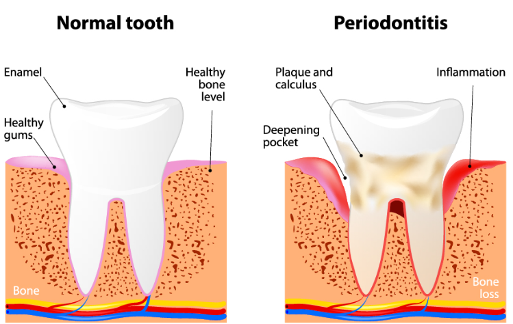
Reason: Deep periodontal pockets make it difficult for the scanner’s light to reach the bottom, and the scanner head has limited angles.
Result: The bottom of the pocket and the root surface may not be captured, creating “blind spots.”
Clinical Impact: Severe missing points can prevent a complete digital impression, affecting restoration design or diagnosis.
2️⃣ Subgingival Margins

-
Reason: The crown or restoration margin is located below the gum line, which blocks the scanner’s light.
-
Result: The margin contour may not be fully captured, resulting in gaps or incomplete edges.
-
Clinical Impact: Poorly defined restoration margins can lead to poor fit, marginal leakage, or secondary caries.
3️⃣
Distal Surfaces of Posterior Teeth
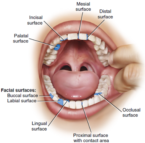
Reason: Posterior teeth are located at the back of the mouth, leaving limited space for the scanner and increasing the risk of interference from adjacent teeth.
Result: Distal surfaces are prone to missing scan data or slight misalignment.
Clinical Impact: Incomplete contact or occlusal information may affect occlusion design and neighboring tooth contacts.
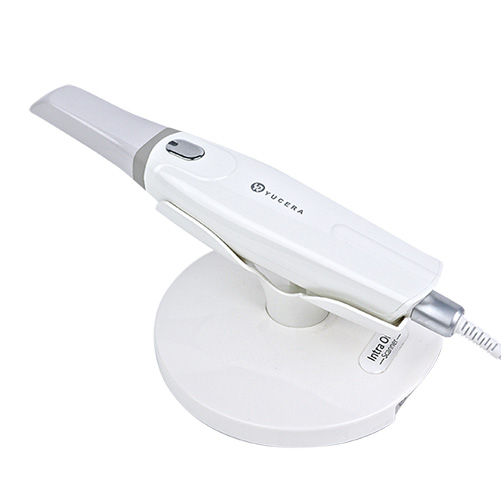
Why the YRC‑S05 Excels at Scanning Challenging Areas
The YRC‑S05 is designed to handle complex intraoral regions such as subgingival margins, distal surfaces of posterior teeth, interproximal spaces, and deep periodontal pockets, thanks to its narrow depth-of-field and high-frame-rate imaging system.
-
Deep Periodontal Pockets & Subgingival Margins: The compact scanner tip, combined with the narrow depth-of-field imaging, allows for flexible angulation after gingival retraction, capturing cervical and subgingival details with high precision.
-
Distal Surfaces of Posterior Teeth: Lightweight design and flexible cabling enable clinicians to easily adjust the scanner angle, minimizing obstruction and improving access to distal regions.
-
Common Missing Points: Data loss is most often caused by saliva interference, limited angulation, or soft tissue movement. The YRC‑S05’s high-frame-rate capture helps reduce these issues, ensuring more complete scans.
Solutions for Common Missing Scan Points
When encountering missing scan points, the following strategies can be applied:
-
Keep the area dry and isolated: Minimize saliva interference and reflective surfaces to ensure optimal optical scanning.
-
Use a light-touch scanning technique: Avoid excessive soft tissue compression that can distort the scan.
-
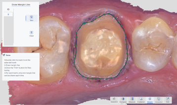
Manually rescan missing areas: Utilize software features for localized rescanning and point cloud repair to complete incomplete data.
-
Use gingival retraction when necessary: Enhance visibility and clarity of subgingival margins.
By applying these measures, the YRC‑S05 achieves a high first-pass scan success rate in routine restorative cases, fully meeting clinical requirements.


 中 文
中 文